Ganglion is a small cystic swelling (not cancer), 2 to 3 cm. It originated in the cavity of the joint or tendon sheath, usually in the wrist but sometimes on foot or knee. From the standpoint of pathology, ganglion is occupied by a wall of fibrous collagen and slimy liquid. Considered to arise as a result of local myxoid degeneration of connective tissue or caused by trauma, such as sprains. Ganglion is usually painless and go, but the treatment.
12/10/2012
Rhabdomyosarcoma
Rhabdomyosarcoma is a fast growing and highly malignant tumor striated muscle. We can identify three distinct clinical and histologic: 1) pleomorphic adults (about 15%), which means it can take different forms, 2) embryonic alveolar (45%), 3) botrioideo embronario (40%). Rhabdomyosarcoma is generally aggressive tumors, early stage widespread, with metastases in the lungs, pleura, mediastinum and pericardium.
Pleomorphic rhabdomyosarcoma occur in men and women, usually between 30 and 40 years. Attacks most commonly the lower extremities, usually the thigh muscles, such as quadriceps, adductor and semimembranosus. The tumor grows quickly and can reach even 25 cm in diameter. The tumor is located deep in the muscles and soft tissues are composed of grayish red meat and fish-like, with a focus of necrosis and hemorrhage. Because these tumors are pleomorphic, with light can be observed microscopically different types of cells, the cells are sometimes racket prolongations protoplasm unique and long, sometimes giant cells with peripheral vacuoles oval band separated by a thin cytoplasm (cell cobweb), but be- liver cell layer usually clearly distinguished from variable dimenciones.
Embryo alveolar rhabdomyosarcoma occurs almost exclusively in children and usually appears at the upper or lower extremities, but sometimes involving the trunk. Macroscopic rhabdomyosarcoma has aspects similar to adults, but rarely reach such large dimensions. In a histological study warns cellular round to oval shape, and the similarity suggests occasional slightly elongated muscle cells mature. The cells are arranged in small nests or rosette, separated by fibrous stroma interlace, frame holds some resemblance to images of lung alveolar cells and alveolar cavity neoplastic occupy, provide a name for this type of tumor.
Botrioideos embryonal rhabdomyosarcomas usually occurs in children and young adults and attacked several sites, for example, genitourinary, biliary tract and orbit. Skeletal muscle is rarely attacked.
Chlorosis (female)
Chlorosis is a disease that occurs in women during the developmental maturity, which consists exclusively of decline in the proportion of hemoglobin in the blood. Chlorosis is widespread among young women in the late nineteenth century. Perhaps chlorotic condition starts very early during embryonic life or during early childhood, but it occurs in young people 12-18 years and associated with ovarian and menstrual cycles. What is striking in this patient was pale, sounding completely white, and ease of fatigue, the lips acquire grayish and the tongue has a bluish gray color.
Symptoms include fatigue and tiredness a bit of effort, the weight of the leg, dyspnea when climbing stairs, scintillating scotoma, headache, cold hands and feet, and menstrual disorders may be amenorrhea, vaginal discharge, and constipation. In more severe cases of chlorosis occurs relative lack of mitral and tricuspid valves. Changes in the characteristics present chlorosis CBC: real hyperchromic anemia, which decrease the proportion of hemoglobin in the blood. The number of erythrocytes is usually normal, but occasionally the upper and lower figures, but the most important change is the proportion of low hemoglobin, which usually varies between 70 and 15%. Therefore, also in the globular value, the amount is less than 0.7, generally varies between 0.5 and 0.6.
In colored extensions poikilocytosis surprising and above all great pale red blood cells. In preparation stained erythrocytes observed some polychromatophils, while in chlorosis, not usually found red blood cells with basophilic granulation. Chlorosis is a chronic condition that can last for several years, more than eight, but the direction is usually seen, usually associated with the treatment and the time of year, it becomes visible annoyance to early spring and early autumn improvement.
Amyloid Substance
Material amyloid (or amyloid) is an abnormal protein substance deposited between the cells of many tissues and organs in dferentes clinical situation. Electron microscopy showed amyloid fibrils as arborescent limited length and a width of approximately 7.5 to 10 nm. These fibrils may occur singly, in groups fasikula lateral or woven mesh. The X-ray and infrared spectroscopy cristografía shows a picture of the characteristics of the so-called "shaping" ß.
We have identified two main chemical classes of amyloid substance: the first is called immunoglobulin light chain AL (amyloid light chain), the second consists of non-immunoglobulin protein AA (amyloid-related protein). This protein is different antigens and stored in different clinical circumstances.
We have identified two main chemical classes of amyloid substance: the first is called immunoglobulin light chain AL (amyloid light chain), the second consists of non-immunoglobulin protein AA (amyloid-related protein). This protein is different antigens and stored in different clinical circumstances.
Myotonic Dystrophy
Myotonic dystrophy, also called Curschmann and Steiner disease is a condition in which muscle atrophy associated with myopathy Thomsen type myotonia and hereditary endocrine disorders. Myotonic dystrophy is a hereditary and family, are more common in men than in women, and the observation of a rare degenerative. Started between 20 and 30 years. The patients had a family history of properties such as high infant mortality, many abortion, sterility, cataracts the beginning of each successive generation.
In myotonic dystrophy, the affected muscles showed a gradual loss of longitudinal and transverse striation and infiltration of the muscle fibers and fat drops of nuclear proliferation. While in the nervous system have been checked degenerative lesions in the diencephalon, and the vegetative nucleus splanchnic nerve, lateral and anterior horn of the spinal cord and posterior. Muscle atrophy that occurs in this disease occurs in the neck, face and distal limbs, myopathy kind uncontracted fibrils.
The condition occurs in myotonic hand, sternocleidomastoid muscle and eyelid. Endocrine and dystrophic signs of premature baldness comprise, eye cataracts, testicular atrophy, mumps, kyphosis or lordosis, with or without hypothyroidism, hypogonadism. The prognosis is not good, as the disease progresses slowly and patients end up as myopathy.
Hair Cells (Ear)
Hair cells sensory cells that are part of the organ of Corti is located in the cochlea in the inner ear. Extension has a very thin, hair-shaped, estereocilias called, stand on the middle level are in contact with endolymph. The function of the hair cells is to convert sound vibrations into nerve impulses that travel to the occipital lobe of the brain via the auditory nerve.


Organ of Corti
Spiral structure of the organ of Corti is formed by the hair cells and supporting neuroepithelial cells. Lying on the basement membrane in the bottom half of the scale, along the cochlea of the inner ear canal, forming a spiral. One can say that the organ of Corti is the auditory center, because it is a place where sound waves are converted into pulses nervisos.
 Vertical cut the cochlea which houses the organ of Corti

12/09/2012
Hemosiderosis
Hemosiderosis calls for storing large amounts of hemosiderin in the organ. Hemosiderin is a protein that stores iron in the body, is a derivative of hemoglobin released during hemolysis (destruction of erythrocytes). Hemosiderosis be recognized macroscopically and in the liver, spleen, pancreas, lymph nodes, etc., ochraceous color. Given to these organs. Histology mainly hemosiderin granules in the cells of the reticuloendothelial system, but also in liver cells in the epithelial cells uriniferous tubes (nephrons, etc.).
Hemosiderosis can be observed as a result of repeated blood transfusions are very broad (transfusional Siderosis) and diseases accompanied by intense blood damage, especially in pernicious anemia. In the lungs also saw marked hemosiderosis, as a result of stasis in the smaller circle has taken a lot of small volume bleeding. Alveolar epithelial then phagocytosed red blood cells to form hemosiderin. When they were expelled in sputum, which is the so-called cardiac cells (cell lesions of the valve), the process is particularly seen in patients with heart valve lesions (mitral valve disease).
Hemosiderosis can be observed as a result of repeated blood transfusions are very broad (transfusional Siderosis) and diseases accompanied by intense blood damage, especially in pernicious anemia. In the lungs also saw marked hemosiderosis, as a result of stasis in the smaller circle has taken a lot of small volume bleeding. Alveolar epithelial then phagocytosed red blood cells to form hemosiderin. When they were expelled in sputum, which is the so-called cardiac cells (cell lesions of the valve), the process is particularly seen in patients with heart valve lesions (mitral valve disease).
Cochlea
The cochlea is the auditory portion of the inner ear, because it is the organ of Corti. It has a spiral structure, with the emergence of a small spiral, and embedded in the temporal bone. The cochlea is divided into three tubular cavity running the spiral staircase: the scala vestibuli, scala media, and scala tympani, the first and the last one is filled with a liquid called perilimfe, but scala media is filled with endolymph, the other fluid types. Vestibuli The scala and scala tympani are connected at the top of the cochlea (the snail), a type of ship called helicotrema communicate. Organ of Corti, which contains neuroepithelial hair cells, the vibrations are transmitted to the cochlea with the bracket through fenestra ovalis (oval window), into nerve impulses that are picked up by the auditory nerve.


12/08/2012
Soft Tissue Fibrosarcoma
Soft tissue fibrosarcoma tumor was malignant tissue. These tumors occur for some time with the good looks fool, which contrasts with the real structure with a large tendency for late recurrence and generalizations. Therefore the interest of early detection. These fibroids develop in filling network and support from all parts of the body: partition and fascias involve muscles, nerves and vessels, showed marked propensity for the extremities. Originally presented as a solitary tumor, the volume varies from size of a walnut to the size of a small orange, but eventually reach a much larger dimension.
In the surgical specimen, the soft tissue fibrosarcomas were packed solid mass. However, the envelope, which is constant, there are barriers that isolate different conjunctiva but is formed by the surface layer of the same tumor, level and strong. They have a very encuenta data, as described excision successive relapse and need extensive subcapsular tumor excision. Mass itself, the court is homogeneous, firm, fibrous appearance and a matte white or grayish white. It consists of connective fusiform cells with large nuclei, without proper guidance, but tend to be arranged around the blood vessels in the wall where they belong. The ship was disorganized, branched, of different calibres.
Recurrence-law, where, after a simple excision, is characteristic of the soft fibroma, a deceptive ease of removal, but after a variable period of full years, almost always new tumor appears under the surgical scar. This tumor recurrence showed a greater tendency to proliferate, as if the transaction had the potential latent elevated her virulent. Thus, the way to the root of the groin, leg or armpit. Therefore it is said, with a sense of mnemonic, this tumor has a "geotropism" negative. The tumor again and again. Progress of the disease and amputations imposed pathology report in the end, the only way to prolong life for some time.
Rhabdomyoma
Rhabdomyoma is a benign tumor composed of striated muscle fibers. It's big and rich fibrils in the cytoplasm of the cell membrane is drawn, which is fixed to it only at some point, when practicing the entry paraffin. Consequently blisters cytoplasmic core center, which emits a thin extension comparable to the spider legs. When developing myocardium reached Rhabdomyomas measuring 2 cm in diameter and were observed in combination with brain lesions (TSC) and the kidneys.
Tumor Cells and Tissue Matrix
Tumor cells can differentiate stem cells that come in almost all important manifestation, but also can preserve many of their properties. As the way we understand the important manifestation, a particular function, metabolism, trasplantabilidad, etc., Image of different species is highly polymorphic tumor.
A) With respect to morphology, tumor cells often retain all the morphological characteristics of their ancestors, and at other times they do not like it at all. Typically, a slow-growing tumor that is structurally most similar to normal tissue (tumor ripe), while growing rapidly have little morphological peculiarities. They remind us of the cells and tissues of the embryo, are also growing rapidly, although it is understood that they are not embryonic cells. In this case too immature and tumor cell dedifferentiation (anoplasia).
B) What is the function of specific tumor tissue is closely related to the same morphological characteristics: the higher the similarity of morphological tumor to normal tissue, the more likely it is also mimic certain functions. This refers primarily to a large number of tumors of endocrine organs. Anyway, there is a parallel force between tumor differentiation and function. In adult tumors usually have a specific function.
C) tumor cells, such as the previous array are affected by specific substances from the organism itself. In the same way that normal prostate or breast tissue responds to hormones, certain tumor tissues before reacting hormónicas excitations as well, and this has been used in therapy.
D) Another important point is that related to the metabolism of tumor cells. All tumors, especially rapidly growing epithelium, behave similarly to embryonic tissue or injured tissue, which is formed at the expense of glucose lactic acid even in the presence of oxygen.
Reissner Membrane
Reissner membrane is a thin membrane that separates two of the three internal parts of the cochlea: medium-sized vestibular scale. It consists of two layers of flattened epithelial tissue, separated by a basal lamina. The upper layer of Reissner's membrane epithelium at the base of the scala vestibuli, which is immersed in a fluid called perilimfe, while the bottom layer, on the roof of the scala media is impregnated endolymph fluid filled cochlea scala media.


Labyrinth
The inner ear is the part of the ear is in the temporal bone. It is composed of three parts: the cochlea, vestibule and semicircular canals. It is a center for hearing and balance. The inner ear is connected to the middle ear through the bracket, which transmits sound vibrations to the cochlea to the ear drum are collected via the oval window. Thanks to the internal fluid cochlea (perilimfe and endolymph), acoustic vibrations are converted into acoustic waves, which are processed, in turn, into nerve impulses by the organ of Corti, the last, which is located in the cochlea scala media, contains hair cells that make this possible . Nerve impulses generated by the inner ear that are sent to the occipital lobe of the brain via the auditory nerve.


Tumor (Concept and General)
Why tumors grow? Tumors grow because their cells multiply constantly. Therefore, the formation, autonomous and independent organization that is not related to the body and is not of itself rather than in relation to nutrition. A tumor on the body behaves as something strange: the cost of their lives as well as parasites, taking from it the necessary nutrients. Growth is generally limited, as only a few cases up to the park. The tumor, therefore, infinite growth neoformations autonomous, ie not connected to the body in an organic-based. The core of this definition is precisely the autonomy of tumor formation.
All the cells of our bodies can grow with tumor phenotypes, albeit in very different degrees. So, for example, connective tissue and epithelial tumors are often, if not go to the nerve cells. Many tumors formed only by a cloth, for example, joints, cartilage or muscle, the other two networks, for example, epithelial tissue, the other with a variety of networks. In the latter case, the different species of cells in a tumor can be set as they would in normal circumstances. Then it is easy to identify its origin. But they also can grow differently: the interstitial cells forming substances can usually proliferate in tumors without training them. Established tumors, and then, only with the cells and corresponding tissue from embryos in difficult circumstances to recognize this source.
Conceptual distinction made here between self-neoplastic cell growth in embryos regeneration, and hyperplasia, especially compared to the neoplasm tumor cells, can be implemented with such precision in all cases.
12/07/2012
Virus
Virus is the formation or structure that consists of a single strand of nucleic acid that is involved in protein coat. Most of them are visible only with an electron microscope. There is a virus that can not be displayed correctly. This virus is not technically considered living organisms because of lack of biological precesos as metabolism and respiration, and can not be played by itself, but requires a host cell to reproduce. Therefore, it is an obligate parasite of the cell.
When a virus invades a cell was first fixed specific receptors on the cell membrane, which has an affinity, to destroy penetrate inside the cell. After that, you can not prove their existence in any way to the top for a while. It is concluded that disintegrate into its constituent elements, which was multiplied by the metabolic processes of cells to join later in greater numbers and become the basic cells. The virus can pass through the two daughter cells when a cell divides, spreading defined by this mechanism.
When a virus invades a cell was first fixed specific receptors on the cell membrane, which has an affinity, to destroy penetrate inside the cell. After that, you can not prove their existence in any way to the top for a while. It is concluded that disintegrate into its constituent elements, which was multiplied by the metabolic processes of cells to join later in greater numbers and become the basic cells. The virus can pass through the two daughter cells when a cell divides, spreading defined by this mechanism.
Semicircular Canal
Semicircular canals are three small tubes, closed loop projecting from the front of the inner ear, three semicircular shape: the previous channel, horizontal and then. Each has a diameter of about 0.8 mm and is filled with a fluid called endolymph. The role of the semicircular canals is to maintain balance and spatial orientation, the sensory cells of the hair, which is related to the vestibular nerve fibers that transmit impulses to the brain via the auditory nerve.


Dermatropos Virus
Virus Dermotropos are those that colonize the skin cells of the epidermis, has as input the body through the pharyngeal mucosa and that usually causes coughing slightly. From it through blood and dermotropo virus reaches the skin, where the occupation. We can show the presence in it called Guarnieri corpuscles were found in the cytoplasm and culture is a small virus. The elementary body, which are relatively large and in accordance with the same virus, known since 1907. Viruses cause infectious dermatropo eruptions such as measles, chicken pox, measles and fever blisters.
After the colonization of the virus in epithelial cells first develop papules, which by damage to the basal layer of the epidermis is converted into vesicles, which soon became turbid water content and become purulent, thus becoming pustules formed. When they dry, crust, which despite all the superficial scars. Both vesicles and crust is a highly contagious virus for their content. When inoculated into rabbit corneas (Paul test) also develops in the epithelial cells Guarneri blood cells. Dermotropo virus leaves patients permanently overcome immune diseases.
After the colonization of the virus in epithelial cells first develop papules, which by damage to the basal layer of the epidermis is converted into vesicles, which soon became turbid water content and become purulent, thus becoming pustules formed. When they dry, crust, which despite all the superficial scars. Both vesicles and crust is a highly contagious virus for their content. When inoculated into rabbit corneas (Paul test) also develops in the epithelial cells Guarneri blood cells. Dermotropo virus leaves patients permanently overcome immune diseases.
Staphylococci
Staphylococci are bacteria cocáceas group. This gram-positive cocci, measuring between 0.5 and 1.5 microns in diameter, forming a clustered multiply accumulation. According to their behavior on solid culture media, are divided into several subgroups: 1) Staphylococcus aureus, which form colonies golden yellow, 2) citreus staphylococci, with lemon yellow, 3) Staph color white dawn. On our skin usually staphylococci, where he underwent a saprophytic life. But sometimes penetrate the tissue of the wound or natural opening in the skin, such as hair follicles or glands sudorípidas, then act as pathogens. The most frightening thing is Staphylococcus aureus, while the disease is rare dawn.
Staphylococci produce a poison called hemolysins which dissolves red blood cells and causes tissue coagulation necrosis (coagulase). Staphylococci are generally distributed to the network a bit, which only produces local lesions characterized by necrosis and pus formation, a purulent infection. They are responsible for ulcers. In open fractures can cause pyogenic infections, and osteomyelitis. In newborns can cause exfoliative dermatitis Ritter.
Staphylococci produce a poison called hemolysins which dissolves red blood cells and causes tissue coagulation necrosis (coagulase). Staphylococci are generally distributed to the network a bit, which only produces local lesions characterized by necrosis and pus formation, a purulent infection. They are responsible for ulcers. In open fractures can cause pyogenic infections, and osteomyelitis. In newborns can cause exfoliative dermatitis Ritter.
Streptococcus
Streptococcus is a spherical bacterium (coccus) which on division (fission) chain formed. There are several types of streptococci and group them into three groups: alpha-hemolytic, beta-hemolytic and non-hemolytic-range, based on the action or the effect that the red blood cells (erythrocytes). Secrete toxins that cause infectious diseases such as angina, erysipelas, scarlet fever otitis, and pneumonia, as well as autoinumitaria disease: rheumatic fever.
The amount of toxins released by these bacteria are important in the diagnosis and treatment will lead to disease. Alpha and beta hemolytic streptococcus form of a hemolysin which attack and dissolve the red blood cells, the body responds to this attack called antihemolisina antibodies. The demonstration of the presence in the blood antihemolisina an important diagnostic tool in the recognition of the disease is not proved by other processes. Exotoxin produced by alpha-hemolytic streptococci produces paralysis reddish blood vessels, for example, scarlet fever. In erysipelas, found beta-hemolytic streptococci, which usually has penetrated through a small hole in the skin of the face, spreading to the lymph nodes examined and crannies.
In chronic streptococcal infection, allergic reaction occupies the foreground of the fabric, while the same bacteria that settle on a focus that is often not easy to find, for example, granulomas in the root of the tooth. Natural focal infections persistent toxins into the body and sensitive. Thus, in various organs develop streptococcal lesions do not contain any, but in the end, is because of the action. In a wide circle of streptococcal disease should include glomerulonephritis, arthritis and rheumatic fever.
Hemosiderosis
Hemosiderosis calls for storing large amounts of hemosiderin in the organ. Hemosiderin is a protein that stores iron in the body, is a derivative of hemoglobin released during hemolysis (destruction of erythrocytes). Hemosiderosis be recognized macroscopically and in the liver, spleen, pancreas, lymph nodes, etc., ochraceous color. Given to these organs. Histology mainly hemosiderin granules in the cells of the reticuloendothelial system, but also in liver cells in the epithelial cells uriniferous tubes (nephrons, etc.).
Hemosiderosis can be observed as a result of repeated blood transfusions are very broad (transfusional Siderosis) and diseases accompanied by intense blood damage, especially in pernicious anemia. In the lungs also saw marked hemosiderosis, as a result of stasis in the smaller circle has taken a lot of small volume bleeding. Alveolar epithelial then phagocytosed red blood cells to form hemosiderin. When they were expelled in sputum, which is the so-called cardiac cells (cell lesions of the valve), the process is particularly seen in patients with heart valve lesions (mitral valve disease).
Hemosiderosis can be observed as a result of repeated blood transfusions are very broad (transfusional Siderosis) and diseases accompanied by intense blood damage, especially in pernicious anemia. In the lungs also saw marked hemosiderosis, as a result of stasis in the smaller circle has taken a lot of small volume bleeding. Alveolar epithelial then phagocytosed red blood cells to form hemosiderin. When they were expelled in sputum, which is the so-called cardiac cells (cell lesions of the valve), the process is particularly seen in patients with heart valve lesions (mitral valve disease).
Pneumococcus
The pneumococcus (Streptococcus pneumoniae) is a spherical bacterium, gram-positive, non-mobility itself, and which cause pneumonia, inflammation of the facial sinuses (sinusitis), and meningitis. Pneumococcus is provided in pairs, ie Diplococcus, as streptococcus, forming chains also multiplied in liquid culture media, but is characterized by sensitivity to bile salts. They also have a mucous capsule containing carbohydrates, especially antigenicity. Using an appropriate immune serum, resulting in swelling of the capsule can diferenicar pneumococci are four main types: type I, II, III and IV.
Injuries caused by a very dangerous type III, characterized by unusually rich mucous exudates which are not formed by the cells of the body, but by the pneumococcus. Type IV is divided into several subgroups. Other pneumococci produce fibrin-rich exudate leukocytes. Pneumococci was a regular guest of the respiratory tract and upper mouth. Although lung infections occur, they can, through the blood to distant organs, such as leptomeninges, as well as for children without a safety peritoneum can be shown by the gateway.
Injuries caused by a very dangerous type III, characterized by unusually rich mucous exudates which are not formed by the cells of the body, but by the pneumococcus. Type IV is divided into several subgroups. Other pneumococci produce fibrin-rich exudate leukocytes. Pneumococci was a regular guest of the respiratory tract and upper mouth. Although lung infections occur, they can, through the blood to distant organs, such as leptomeninges, as well as for children without a safety peritoneum can be shown by the gateway.
Chickenpox
Chickenpox is a rash illness of acute, infectious, fever. The rash consists of small particles, the part that turns into vesicles dried without flowing or ulceration. This disease affects children and is caused by the varicella zoster virus. It is highly contagious and spreads through the air exhaled by the patient, without the need for direct contact with it. Chickenpox begins suddenly, without prodromal manifestations with fever, generalemente of 38 ° (often less than 39 °). The rash usually appears first on the face and scalp, and then served on the trunk and extremities. After several hours, the stains become transparent vesicles surrounded by hyperemic halo.
pathology
Bile, which is a measure, lentil unilokular not multilocular (so not emptied the entire contents immediately vesicles to puncionarla) but. Serum vesicles contain some leukocytes and staphylococci often. After one or two days, where the temperature is gradually decreased, the vesicles dried and formed a small central depression. Drying gallbladder verified after becoming the first item and then cloudy yellowish, dry scab forms, which comes after eight to ten days.
treatment
Because chicken pox is caused by a virus, antibiotics are not effective. Therefore, the limited treatment to relieve symptoms. The prognosis is good in 99% of cases and leaves immune diseases.
Hemochromatosis
Hemochromatosis is a recessive inherited condition characterized by changes in iron metabolism, leading to large accumulation in the body tissues of iron-containing protein called hemosiderin, which is a byproduct of hemoglobin produced hemolysis. Therefore, hemochromatosis causes excessive iron in the body. Consequently, there are also tanks lipid pigment granules called lipofuscin, in intestinal smooth muscle found some excessive lipofuscin and melanin in the skin, so it has a chocolate-brown. Symptoms of hemochromatosis are: weight loss, fatigue and general weakness, heart palpitations, agitation, etc..
treatment
For patients suffering from hemochromatosis is an iron overaccumulation treated felbotomías practicándoseles, which were removed by the blood of patients, in the same way that blood taken from blood donors. This is done once or twice a week. Phlebotomy accompanied by a diet low in iron.
treatment
For patients suffering from hemochromatosis is an iron overaccumulation treated felbotomías practicándoseles, which were removed by the blood of patients, in the same way that blood taken from blood donors. This is done once or twice a week. Phlebotomy accompanied by a diet low in iron.
Lipofuscin
The lipofuscin pigments are lipid (fat) and yellowish brown granular, without iron. It is formed as a result of the oxidation of lipids and protein residues. Lipofuscin is found in many tissues and organs (liver, kidney, heart muscle, adrenal glands, etc.). Usually accumulates with age in the lysosomes of cells, and the accumulation of risk factors in the epithelium of the retina-related macular degeneration age, because it can damage the retinal photoreceptor cells.
Uric Acid (Purpose and Location)
Uric acid is the end product of purine metabolism and protein nitrogen. Formed as an intermediate product of normal metabolism and is eliminated by the kidneys that are mostly muscle. When there is an increase in pathological acid formation and decreased their removal has a direct effect on the tissues of the body. Salt of uric acid stored in individual tissues in the form of monosodium urate, which is a sign of disease anatomical characteristics we call drop. Preferred location is uric acid cartilage and joint capsule.
Vein deposits also form small nodules or tophi, in the ear cartilage, tendon sheath and bag in the mucosa. Some crystals, gathered in bundles, developed granulation tissue with foreign body giant cells. As the cause of gout should be taken into account on the one hand certain adverse exogenous factors (poor diet rich in purines), and, second, constitutional factors.

Vein deposits also form small nodules or tophi, in the ear cartilage, tendon sheath and bag in the mucosa. Some crystals, gathered in bundles, developed granulation tissue with foreign body giant cells. As the cause of gout should be taken into account on the one hand certain adverse exogenous factors (poor diet rich in purines), and, second, constitutional factors.

Uric acid is the chemical formula
Gaucher Disease
Gaucher disease is a rare disease characterized by the deposition of fatty molecules called cerebrosides, in cells of the reticuloendothelial system and can affect various organs, including the liver, spleen, bones and central nervous system. Gaucher disease is hereditary, irregular dominant character, and affects infants, children and adolescents. This lipid accumulation due to absence of the enzyme called glucocerebrosidase, which acts with cerebrosides decay, thereby preventing precipitation or accumulation of these cells. The lack of this enzyme is caused by mutations in a gene located on chromosome 1. This condition was first described by French physician Philippe Gaucher in 1882.
The main symptoms of Gaucher disease is an enlarged liver and spleen, bone weakness (with severe pain in the bones), nervous system damage, and anemia. Effective treatment involves the application of Gaucher disease, endovenous, of the enzyme glucocerebrosidase, which is obtained from the placenta or recombinant DNA imiglucerasas.
There are three recognized forms of the disease: type I, II and III. Type I is the most common form, occurs at any age and the symptoms are more docile and attacks the nervous system. The type II only occurs in children and attacks the nervous system. The type III usually begins in childhood and also showed symptoms of Type I and II, and come in an average of one per thousand births cicuenta.
Middle Ear
The middle ear is the part of the auditory system that lies between the outer ear and the inner ear. It consists of the eardrum and a chain of three tiny bones that transmit vibrations from the eardrum. These bones are: hammer, anvil and stirrup, united by cartilage. The eardrum is a thin membrane composed of translucent connective tissue and epithelial acoustic vibrations picked up from the outside and sends the hammer, anvil and stirrup, which in turn sends them to the inner ear through the oval window. The function of the middle ear is, therefore, receive and transmit sound waves from the outer to the cochlea, located in the inner ear.


12/05/2012
Vestibular Nerve
Vestibular nerve is one of the two branches that form the auditory nerve. It extends from the semicircular canals, through the vestibular ganglion, to the junction with the cochlear nerve to form the auditory nerve or cranial nerve VIII. Vestibular nerve sends nerve impulses related to balance and spatial orientation. The fibers of the vestibular nerve axons synapse with neurons in the vestibular nuclei annular bulge located on the wall of the fourth ventricle.
Pfaundler-Hurler Disease
Pfaundler-Hurler disease, or mucopolysaccharidosis gargoylism also called, is a hereditary disease characterized by mental retardation, amaurosis (blindness), deafness, impaired development of the skeleton that causes dwarfism tarnish. Pfaundler-Hurler disease is due to disturbances in the metabolism of phosphatides and accumulation of mucopolysaccharides in the absence of a lysosomal enzyme (alpha-L iduronidase), which is responsible for the degradation of mucopolysaccharides in the lysosomes of cells. Treatment: Application of the missing enzyme, bone marrow transplantation and cord blood.
Niemann-Pick
Niemann-Pick disease is caused by a disturbance in the metabolism of phosphatides (phospholipids), characterized by the deposition of sphingomyelin in the reticuloendothelial system cells and lymph nodes, but also the cells and the endothelial tissue. Niemann-Pick disease is inherited in a recessive and seen mainly in children. Typical signs of the disease are gastrointestinal disorders, anemia, hypertrophy and abnormalities of the liver and spleen, as well as neurological deficits. Typically, children who suffer from this disease do not survive three years.
Auditory Nerve
Auditory nerve, or vestibulocochlear nerve is the eighth nerve of the twelve pairs of cranial nerves. It comes from a combination of vestibular and cochlear nerve in the inner ear. Therefore, the auditory nerve is composed of fibers that transmit nerve impulses from the balance and space, which is derived from the bipolar cells in the vestibular ganglion contact with the semicircular canals, and fiber that comes from the sense of hearing in the body of Corti in the cochlea. Auditory nerve (VIII) sends nerve impulses to the brain via the spinal accipital lobe.
Sphingomyelin
Sphingomyelin is a phospholipid composed of sphingosine, choline, phosphoric acid, and fatty acids. It is located in the cell membrane of neurons of the nervous system, forms part of the myelin covering the axon. It is believed that sphingomyelin plays a role in signal transduction (extracellular signals).
Hypoglossal Nerve
Hypoglossal nerve is the twelfth cranial nerve. Needs of the tongue muscles. It comes from the core of their motor neurons form in the spinal anterolateral sulcus between the olive and the pyramid. When the axons of these neurons join to form the common troco nerve emerges from the skull through the hypoglossal canal. Then XII cranial arc and is divided into terminal branches, which innervate various muscles tongue.
Collateral Circulation in The Arteries
Occlusion of the artery ischemia occurs not only in cases where it is impossible for the blood of the arrival of other ways. But too often there is the possibility that the blood out another way to areas blocked artery, and often enough, and this is known as collateral circulation in the arteries. It often happens that a clogged artery branches anastomose with other neighbors, then both carry oxygenated blood to the area which had been removed from circulation. This is known as anastomosis, or collateral line, tying the branches, and collateral circulation built this way.
Collateral circulation varies from case to case. Sometimes quite from scratch and others (initially) solid. Let's first collateral circulation and there are usually between the radial and ulnar arteries, through the palmar arch. If the radial artery is blocked, the blood comes to the palm of the ulnar-at first, so that the blood reaches approximately half of the previous. But soon the blood flow through the ulnar faster than normal, so the hand will reach more of the same.
The opposite occurs when a narrow anastomosis that, despite the acceleration of blood flow and dilation of vessels immediately, it is not a sufficient amount of blood. This raises the disorder, may still be compensated. Collateral vessel dilation, which at first is that according to the normal capacity of adaptation, after exceeding the limit. Each element is multiplied walls, and glass, narrow at first, a big glass. Not only that there is a narrow artery anastomosis can experience this kind of growth, but under special conditions capillary tube could also be widened arteries and form on his wall all the characteristic elements of the arterial wall, thus avoiding tissue necrosis.
Collateral circulation can, therefore, even in a difficult pose but its development is much more difficult when there is a narrow collateral. However, sometimes the network does not support long prolonged blood deficit. Such as occurs, for example, when the femoral artery is clogged or garter above the starting point of the profunda femoris. Then established collateral circulation through the arterial anastomosis between the hip and femoral arterial muscle, so it is often too slow, then become necrotic foot and part of leg.
12/04/2012
Vestibular Nerve
Vestibular nerve is one of the two branches that form the auditory nerve. It extends from the semicircular canals, through the vestibular ganglion, to the junction with the cochlear nerve to form the auditory nerve or cranial nerve VIII. Vestibular nerve sends nerve impulses related to balance and spatial orientation. The fibers of the vestibular nerve axons synapse with neurons in the vestibular nuclei annular bulge located on the wall of the fourth ventricle.
Pfaundler-Hurler disease
Pfaundler-Hurler disease, or mucopolysaccharidosis gargoylism also called, is a hereditary disease characterized by mental retardation, amaurosis (blindness), deafness, impaired development of the skeleton that causes dwarfism tarnish. Pfaundler-Hurler disease is due to disturbances in the metabolism of phosphatides and accumulation of mucopolysaccharides in the absence of a lysosomal enzyme (alpha-L iduronidase), which is responsible for the degradation of mucopolysaccharides in the lysosomes of cells. Treatment: Application of the missing enzyme, bone marrow transplantation and cord blood.
Niemann-Pick
Niemann-Pick disease is caused by a disturbance in the metabolism of phosphatides (phospholipids), characterized by the deposition of sphingomyelin in the reticuloendothelial system cells and lymph nodes, but also the cells and the endothelial tissue. Niemann-Pick disease is inherited in a recessive and seen mainly in children. Typical signs of the disease are gastrointestinal disorders, anemia, hypertrophy and abnormalities of the liver and spleen, as well as neurological deficits. Typically, children who suffer from this disease do not survive three years.
Auditory Nerve
Auditory nerve, or vestibulocochlear nerve is the eighth nerve of the twelve pairs of cranial nerves. It comes from a combination of vestibular and cochlear nerve in the inner ear. Therefore, the auditory nerve is composed of fibers that transmit nerve impulses from the balance and space, which is derived from the bipolar cells in the vestibular ganglion contact with the semicircular canals, and fiber that comes from the sense of hearing in the body of Corti in the cochlea. Auditory nerve (VIII) sends nerve impulses to the brain via the spinal accipital lobe.
Sphingomyelin
Sphingomyelin is a phospholipid composed of sphingosine, choline, phosphoric acid, and fatty acids. It is located in the cell membrane of neurons of the nervous system, forms part of the myelin covering the axon. It is believed that sphingomyelin plays a role in signal transduction (extracellular signals).
Hypoglossal Nerve
Hypoglossal nerve is the twelfth cranial nerve. Needs of the tongue muscles. It comes from the core of their motor neurons form in the spinal anterolateral sulcus between the olive and the pyramid. When the axons of these neurons join to form the common troco nerve emerges from the skull through the hypoglossal canal. Then XII cranial arc and is divided into terminal branches, which innervate various muscles tongue.
Collateral circulation in the arteries
Occlusion of the artery ischemia occurs not only in cases where it is impossible for the blood of the arrival of other ways. But too often there is the possibility that the blood out another way to areas blocked artery, and often enough, and this is known as collateral circulation in the arteries. It often happens that a clogged artery branches anastomose with other neighbors, then both carry oxygenated blood to the area which had been removed from circulation. This is known as anastomosis, or collateral line, tying the branches, and collateral circulation built this way.
Collateral circulation varies from case to case. Sometimes quite from scratch and others (initially) solid. Let's first collateral circulation and there are usually between the radial and ulnar arteries, through the palmar arch. If the radial artery is blocked, the blood comes to the palm of the ulnar-at first, so that the blood reaches approximately half of the previous. But soon the blood flow through the ulnar faster than normal, so the hand will reach more of the same.
The opposite occurs when a narrow anastomosis that, despite the acceleration of blood flow and dilation of vessels immediately, there is an adequate amount of blood. This raises the disorder, may still be compensated. Collateral vessel dilation, which at first is that according to the normal capacity of adaptation, after exceeding the limit. Each element is multiplied walls, and glass, narrow at first, a big glass. Not only that there is a narrow artery anastomosis can experience this kind of growth, but under special conditions capillary tube could also be widened arteries and form on his wall all the characteristic elements of the arterial wall, thus avoiding tissue necrosis.
Collateral circulation can, therefore, even in a difficult pose but its development is much more difficult when there is a narrow collateral. However, sometimes the network does not support long prolonged blood deficit. Such as occurs, for example, when the femoral artery is clogged or garter above the starting point of the profunda femoris. Then established collateral circulation through the arterial anastomosis between the hip and femoral arterial muscle, so it is often too slow, then become necrotic foot and part of leg.
12/03/2012
Consequences of Edema
The consequences of edema located mainly in the mechanical obstruction of the affected body organ function. We see, for example, pulmonary edema, alveolar cavity filled with fluid prevents gas exchange through the walls of the alveoli edema aryepiglottic folds (glottis edema) results in narrowing of the respiratory tract and sometimes die by this mechanism, optic nerve edema can cause blindness, the pressure of ascites fluid can compress the inferior vena cava and thus produce blood stagnation in the field of glass roots, accumulation of fluid in the chest compress the lungs and can be dangerous by causing mediastinal shift occasionally.
Whooping Cough
Pertussis, or whooping cough, is a contagious infectious disease that primarily occurs in children, caused by the bacterium Bordetella pertussis. Pertussis is characterized by coughing fits seizures. The incubation period of this disease usually lasts five to fourteen days. The disease is divided into three periods or phases: 1) catarrhal, 2) seizures or paroxysmal, 3) and resolution.
Catarrhal stage may begin tórpidamente, children are cranky, lose their appetites and happening now cold, effusion of mucus, wheezing, hoarseness cough. Cough up the intensity and within two weeks of seizures usually alleges period, with coughing paroxysms where children face a dark red, swollen and there labiros thick and foamy sputum. After the climax, the child was temporarily replaced until the next coughing fit. By moving period of seizure increases the frequency of paroxysms and the number of repetitions and felt by auscultation sibilant rales and rhonchi. Phase seizure does not end in a fast, but through a gradual decrease or resolution period, when children take three to five weeks of paroxysmal cough. In the resolution phase paroxysms stop night and day became less frequent, lasts up to four weeks.
Complications mainly bronchopneumonia, which occurs when inflammatory lesions of the mucosa larynx, trachea, and major bronchi, small bronchi and bronchioles spread. Bronchopneumonia characterized by a sudden rise in temperature, prostration, anorexia, dyspnea and tachycardia.
Treatment: erythromycin, but it is effective when given in the first stage.
Malta Fever
Malta fever, also called Brucellosis is an infectious disease caused by Brucella melitensis micrococci or Bruce, young coconut, gram-negative, with the fast Brownian motion. He was usually spread Malta fever with goats or cows consuming raw milk and its derivatives, and other contaminated food, the gateway of the intestinal bacteria. The disease is found in all temperate regions of the Mediterranean, especially along the coast and islands, but also in other countries were warm.
Malta fever begins after an incubation period of eight to fourteen days, with symptoms including fever, headache and muscle, dyspepsia, etc.. Fever is usually reached 40 and 41 ° C for three to four days, then declined in the week with a morning remission. In chronic cases, brucellosis causes weight loss, decrease in the proportion of hemoglobin in the blood and the number of erythrocytes.
treatment
Prevention by vaccination. Antibiotics such as tetracycline and sulfonamides effective.
Capillariasis heart
The liver is a rare parasitic capillariasis in humans, caused by Capillaria hepatica, a parasitic nematode from 2-10 cm, which lives in the hearts of mice and other mammals. Symptoms consisting of fever, hepatomegaly, and elevated eosinophilia. Capillariasis may resemble liver hepatitis or liver abscess. Pathophysiology is derived by the invasion of the liver by adult worms and eggs, the latter causing granulomas. This is serious if left untreated. The diagnosis is made only by the identification of parasites or eggs in the liver biopsy or autopsy.
The life cycle of Capillaria liver performed when an infected animal dies and liver, eggs contain, decomposes on earth. Also, when carnivores eat the liver of infected animals and remove the eggs with dirt on the ground.
The life cycle of Capillaria liver performed when an infected animal dies and liver, eggs contain, decomposes on earth. Also, when carnivores eat the liver of infected animals and remove the eggs with dirt on the ground.
Diphtheria
Diphtheria is an infectious disease caused by a toxin Loeffler bacillus (Corynebacterium diphtheriae). The most common is diphtheria pharynx, where toxins of bacilli destroy healthy tissue in the upper throat around the tonsils, leading to the formation of a false membrane in the gray color of the mucosa of the same. Symptoms include fever, difficulty breathing and swallowing. Infection occurs microdroplets esparsen through which the air by sneezing or coughing of the patient. When compounded, Diphtheria is a serious disease and can cause inflammation of the myocardium (myocarditis) and damage to the nervous system.
The incubation period is two to seven days. Children are loss of appetite, fever and prostration appears. Vomiting sometimes occur. Face flushed and children over the voice call on breathing, dull noise through the mouth. Tongue dry and covered with fur, to examine the mouth and throat, redness warned intense pharyngeal mucosa and the formation of gray, arranged in spots or bands in the tonsils, quickly assembled and appear as embedded in the mucosa. Pharyngeal lesions develop in a few days. At the beginning of oral disease almost beyond 39 ° and, in many cases, after one week and disappear plaque and produces a gray throat cleared, no internal organ changes occur. However, many may have serious heart complications and nervous system.
treatment
In severe cases, hospitalization is required of patients treated with diphtheria antitoxin and antibiotics.
Mumps Orchitis
Mumps orchitis is known as inflammation of the testis by mumps. Symptoms include pain and testicular inchazón, fever and loss of control. Type orchitis occurs in men in their early teens who recently suffered from goiter. No it is not treated and if you have complications can cause testicular atrophy.
Naegleria Fowleri
Naegleria fowleri is a genus of protozo, which has a life cycle phase and other amoeboid falgelada. Some species Naegleria fowleri is very parasitic and pathogenic, causing primary amoebic meningoencephalitis, which is very serious. Since this is protozo in stagnant pond, lagoon waste and acquired infection swimming in waters contaminated with amoebae.
Naegleria fowleri reproduce by binary fission, faster in warmer temperatures. Having your body core and surrounded by a membrane and move through pseudopodia tranparente. At low temperatures have a tendency to encyst.
Naegleria fowleri enters the central nervous system through the mucous membranes of the nose olfactorias, attacking first the olfactory bulb, and then, to corrode the frontal cortex. Fortunately, these infections are very rare and occur in tropical countries of Asia and northern Australia, where it was discovered in 1965.
Naegleria fowleri enters the central nervous system through the mucous membranes of the nose olfactorias, attacking first the olfactory bulb, and then, to corrode the frontal cortex. Fortunately, these infections are very rare and occur in tropical countries of Asia and northern Australia, where it was discovered in 1965.
12/02/2012
SFA
SFA is one of the two main branches of the femoral artery and the other is the deep femoral artery. Born to adjust where the neck of the femur in the upper leg. Superficial femoral artery have a field parallel to the femur bone and projecting along the route and genicular artery branches down muscle. Irrigate with oxygenated blood to the leg muscles.


Intercostal Neuralgia
Intercostal neuralgia is pain in the chest wall due to inflammation of the intercostal nerves. It can be seen as toxiinfecciosas cause herpes, anemia, consumptive state, and is characterized by pain to the exacerbation of violence, sometimes throbbing sitting on his way from the affected nerve or nerves.
In intercostal neuralgia, pain in the course of the nerve to put pressure on the axillary line height, (points Valleix) paravertebral and parasternal. Intercostal neuralgia is more common on the left than on the right and mainly affects elderly women. Diagnosis should be done carefully so as not to confuse with raduculalgias by the tumor, pleural disease, tabes, pericarditis, epidemics pleurodimia and broken ribs.
Treatment: Anti-inflammatory, vitamin B12.
Knee Down Arteries
Arterial down knee, also called down genicular arteries, the blood vessels that extend from the bottom of the superficial femoral artery. Before reaching the knee, down genicular artery, is divided into two branches: a branch of saphenous and articular branches. It provides oxygenated blood to the muscles and ligaments of the knee.


Tific Bacillus
Tific Bacillus (Bacteria typhi), also known as Salmonella or Eberth bacillus, the bacterium that causes typhoid fever. It is a gram-negative, is about 1 to 2 microns in length and has a rounded tip. Can grow very well in all culture media than alkaline. Tific bacillus people enter through the mouth and invade the body through the lymphatic system in the gastrointestinal tract. Thus, the bacilli reach the mesenteric lymph nodes through lymphatic vessels, passes it to the blood marrow, spleen and bone. Tists bacilli not multiply in the blood, but in the mesenteric lymph nodes, spleen and bone marrow, from which burst into the blood and reach different parts of the body. Secrete endotoxins that cause disease.
Basil was found in a bloody eliminated by the kidneys, indicating its presence in the urine. At the same time pass and colonize besícula bile ducts, from which in turn passed back to the intestine, thus, the maximum cuand proliferation in the gallbladder (third week) can easily be demonstrated in the feces. Some of the bacillus that has penetrated tists blood and body finally gave up, because, after all, free endotoxin contained in the body of bacteria. Toxins are primarily injure parenchymal tissues of different organs, including the myocardium them. In skeletal muscle fibers come strange homogenization, which makes them yellow and brittle.
Basil was found in a bloody eliminated by the kidneys, indicating its presence in the urine. At the same time pass and colonize besícula bile ducts, from which in turn passed back to the intestine, thus, the maximum cuand proliferation in the gallbladder (third week) can easily be demonstrated in the feces. Some of the bacillus that has penetrated tists blood and body finally gave up, because, after all, free endotoxin contained in the body of bacteria. Toxins are primarily injure parenchymal tissues of different organs, including the myocardium them. In skeletal muscle fibers come strange homogenization, which makes them yellow and brittle.
Plague Pneumonia
Plague pneumonia is highly contagious infection, caused by inhaling bacilli expelled by coughing Yersin plague patients with this form. Pulmonary infection with clinically from chills, high fever and paroxysmal cough with bloody sputum expectoration. Occur isolated or confluent bronchopneumonia, usually in the lung lobes simultaneously. With cough bacillus Yersin issued. Death is usually the third or fourth day, when the patient is not treated, with signs of septicemia. Plague pneumonia is a form of pulmonary plague, both caused by the same bacteria, which in turn is transmitted to humans by fleas from rats.
treatment
Before the discovery of antibiotics, especially in the Middle Ages, this used to be a very deadly disease. As an infectious disease caused by bacteria, antibiotics will make a difference.
treatment
Before the discovery of antibiotics, especially in the Middle Ages, this used to be a very deadly disease. As an infectious disease caused by bacteria, antibiotics will make a difference.
Saphenous Artery
Saphenous artery is one of the two branches of the artery that divides down the knee, the other is the articular branch. Within a short distance vertically down along the lower end of the thigh, finishing in three small and short branches wilt slightly below the knee. The saphenous artery anastomosis without the other arteries and contributes very little to irrigation legs and knees.


Lower Limb Arteries
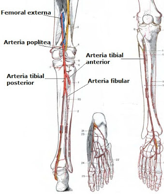
All of the lower limb arteries carry oxygenated blood to, bones muscles and ligaments in the knee, thigh and leg. All this comes from the femoral artery, which in turn is derived from the external iliac artery. Therefore we can say that the femoral artery is the mother of all lower extremity arterial system, which is a continuation of the external iliac, ranging from the level of the inguinal ligament in the pelvis.
After driving a short distance of about 10 to 12 cm, divided into deep femoral artery and femoral external femoral, the first project the following branches: the lateral circumflex femoral artery, a branch off the lateral femoral circumflex and branches of the perforator supplying the femur. In contrast, the external femoral have longer course, supply the muscles of the thighs, and reach the knee continues or becomes the popliteal artery, which passes behind the knee anterior tibial artery divided into and posterior tibial arteries.
The course also has a leg tibialis anterior decreased and vertical runs along the front, between the tibia and fibula in his journey accompanied by anterior tibial vein, and finally, after passing the upper ankle joint, a dorsalis pedis artery. Tibialis posterior, larger caliber than ever, along the back of the foot and provides the following branches: artery fibular (or peroneal), mean arterial plantar and lateral plantar arteries, which carry oxygenated blood to the legs.


Popliteal Artery
Popliteal artery is the main blood vessel that carries oxygenated blood to the legs. At the end of it, was born at the height of the top of the knee, as a continuation of the superficial femoral artery. After traveling about 11 cm vertically along the back of the upper leg and knee, popliteal artery end route is divided into two main branches: the anterior tibial artery and posterior tibial arteries. However, before completing his journey, popliteal project following branches: Sural artery, superior genicular artery, genicular artery lower middle genicular artery.


Anterior tibial artery
A. tibialis anterior is one of the two branches in which the popliteal artery, the other is the posterior tibial artery. Born on the top and back of the leg, just below the knee, and then advanced through the space between the tibia and fibula. Then descend vertically, traveling along the front of the foot parallel to the fibula. When the anterior tibial artery reaches the point ankle joint, renamed dorsalis pedis (or dorsalis pedis).


Cloudy Swelling
Cloudy swelling is a disorder of protein metabolism. Protoplast pellets containing more than usual, and therefore less transparent and appear blurred. At the same time the cell is enlarged, ie, swelling. It's nothing that mitochondrial granular opacity of the material, greatly increasing the volume of water binding. The murky appeared in numerous intoxication, for example, phosphorus and arsenic, as well as by bacterial toxins (diphtheria basil, basil tists, Streptococcus, etc.). The same macro and microscopic images can also develop in organs after death as the body changes. It is also present in the cells of the organ murky been experiencing excessive surgery, for example, the kidney removed after the other.
Cloudy swelling may not be a serious functional impairment of protein metabolism and easily reversible, because one must accept that occurs in many diseases healed without sequelae persistent parenchymal organs. However, when it lasts for a long time, serious injuries can occur, such as adiposity, and ultimately cell death.
Escherichia Coli
Escherichia coli, also known as colibacillus, is one of the most extensive and bacteria live in the intestinal tract in a state of symbiosis. A large negative bacilli, rub-shaped, about 2 microns in length and a diameter of 0.5. Shortly after the birth of man, through the intestinal tract to be constant are the same throughout life. Escherichia coli involved in the disintegration of food, play an important role in the formation of vitamin B12 and K. Basil intestine because it is not a human intestinal saprophyte casual, but human life is possible because of their symbiosis. Intact skin and mucosa which, under normal circumstances, adequate protection against the penetration of Escherichia coli in the network.
However, various modifications of the intestinal mucosa can be made impermeable to germs, whether inflammatory necrosis, mild or injury. Then act as an agent of the colon bacillus flogógenos (which causes inflammation) that infiltrate the intestinal wall and infect the peritoneum. It is clear then that play a role in inflammatory bowel organs and all its attachments. We also found Escherichia coli to inflammation of the pancreas, gallbladder appendix,, together with other intestinal bacteria, and even without that in each case they have been the main agent of the swelling. But sometimes it also penetrate into the bloodstream (sepsis E. coli) and then eliminated by the kidneys, without it being necessary to channel pain or kidney or urinary tract. Only when the urine stagnates in places produce serious injuries. Penetration into the urinary tract can also be done from the outside, especially in women, as long orta urethra.
It is possible to distinguish different types of colon bacilli by serological methods, after seeing that some kind is the agent of infectious enteritis infants sometimes presented as a problem of epidemic in the hospital immediately. In infants found colibacteria well as infectious agents leptomeninges, which arrives via the blood or lymphatic routes. Penetration of the blood can be explained by the greater penetration of the intestinal mucosa baby, to address the same through the lymphatic drag sometimes affect the middle ear.
Posterior Tibial Artery
Tibialis posterior is one of the two branches are divided into the popliteal artery at the end of the trip, the other branch is the anterior tibial artery. Descend vertically, covering the back of the leg, parallel to the tibial nerve and muscle irrigate with oxygenated blood, bones and ligaments of the foot and the plantar foot. At the back of the middle of the foot, the posterior tibial artery branches project important: fibular artery (or peroneal). After reaching the inside of the foot, below the ankle, the posterior tibial artery bifurcates into the lateral plantar artery plantar middle.


Dorsalis Pedis Artery
Dorsalis pedis artery arises from the anterior tibial artery, at the height of the upper ankle joint. Project following branches: lateral and medial tarsal artery, arcuate artery, and deep plantar arteries. Dorsalis pedis artery provides oxygenated blood to the feet and toes.


Saprophyte
In medicine, is a saprophyte microorganisms such as bacteria, fungi or protozoa, which feeds particulate organic or dead cells.
Babinski Sign
Babinski reflex neurological signs characterized by the extension of the toes when the plant is frictioned thereof or rubbed firmly with non-sharp objects. It is normal in children younger than two years but a sign the brain or spinal damage in adults.
Meralgia paresthetica
Meralgia paresthetica, also known as Bernhardt-Roth disease, characterized by pain and burning sensation in the femoral nerve innervation of the skin in the anterolateral thigh. The patient also complained of pruritus. No motor weakness and reflexes are preserved. Meralgia paresthetica can last a long time, there are cases that have lasted more than twenty years. It has been reported more frequently in the police and security agencies, as well as a waitress. That is why it appears that one reason is the tension given to the lateral femoral cutaneous nerve when the patient is standing too long (standing), for a few hours, on a regular basis. Other less common causes are traumatic inflammation, while at other times meralgia paresthetica is a symptom of spinal tumor affecting the corresponding root.
Intercostal nerve paralysis
Intercostal nerve paralysis paralysis of the muscles of the chest and the wall of the stomach. The latter, as lower intercostal injury. Intercostal nerve palsy is a rare observation and neuritis can be caused by malaria, typhoid injuries, diabetes and alcohol, or isolated that one or more nerve damage. Manifestations may be painful or paresthesia in the nerve pathways. Shortly commonly affects breathing, to intervene other muscles innervated by the intercostal not.
Paralysis of the muscles of the abdominal wall, a unilateral deviation determines the navel toward the healthy side, and the repeal of the stomach disappeared temporarily paralyzed last prolapse (prolapse) when the patient coughs or tries to sit down.
Paralysis of the muscles of the abdominal wall, a unilateral deviation determines the navel toward the healthy side, and the repeal of the stomach disappeared temporarily paralyzed last prolapse (prolapse) when the patient coughs or tries to sit down.
Accessory Nerve (XI)
Accessory nerve is the eleventh pair of cranial nerves. It is a motor nerve fibers formed by the cranial (by about 30%.), Originating from the top of the brain stem, and spinal cord (70%), which comes from the top of the bone. These fibers combine to form the accessory nerve (XI), which supplies the trapeze muscles (neck and back) and sternocleidomastoid (neck).
Vitamin A
Vitamin A is a yellow crystalline compound, soluble in fat and is essential in the human diet. Contributing to the development and maintenance of different cell tissues, especially epithelial, mucus and retinal rods. Deficiency that causes hardening and roughening of the skin and mucous membranes, night blindness, and decreased effectiveness of the immune system. Studies in animals and humans have shown that the absence of vitamin A result in impaired synthesis of hemoglobin, causing anemia. When discussing the course of a protective effect against the onset of squamous cell carcinoma of vitamins, for example, skin, bladder and lung.
Vitamin A is present in the normal diet in two ways: as a retinyl ester (retinol), and the beta-carotene (pro-vitamin A). In the first form, retinol, is found in abundance in fish liver oils, especially cod, egg yolks, butter, beef liver of animals, etc., while in the form of beta-carotene is found in yellow vegetables, orange and red, such as carrots, pumpkin , papaya, etc.. However, the most effective absorption and fast are the first, the retinyl ester.
Vitamin A is present in the normal diet in two ways: as a retinyl ester (retinol), and the beta-carotene (pro-vitamin A). In the first form, retinol, is found in abundance in fish liver oils, especially cod, egg yolks, butter, beef liver of animals, etc., while in the form of beta-carotene is found in yellow vegetables, orange and red, such as carrots, pumpkin , papaya, etc.. However, the most effective absorption and fast are the first, the retinyl ester.
Kwashiorkor
The protein-calorie malnutrition occurs in two clinical picture: marasmus and kwashiorkor. Marasmus is a state of malnutrition due to lack of calories. Kwashiorkor dependent protein food insufficiency in spite of adequate caloric intake. Kwashiorkor is a language spoken in Ghana and means "the sickness struck the greater when the child is born", that is, when the older child deprived of breastfeeding and weaning to look at diet, particularly carbohydrates. Children with kwashiorkor presenting general edema, short stature, pale, intestinal disorders, anemia, changes in hair (in pigmentation, tensile strength, and sometimes loosening the roots of alternating bands produce discoloration and pigmentation called "flag sign"). Children may have a misleading appearance of fat-dependent general edema. Another feature of kwashiorkor is hepatomegaly and manifestations Less vitamin.
Total calorie diets lack protein disproportionate sometimes produce a mixture of marasmus and kwashiorkor syndrome. All forms of protein-calorie malnutrition is common in almost all developing countries and underdevelopment in Asia, Africa and Latin America. These countries are losing not only make children more vulnerable to infectious diseases, but once contracted can be fatal, because their immune systems are not functioning properly due to the state of malnutrition. A child with kwashiorkor who have contracted an infectious disease at high risk of developing bacterial septicemia.
Changes in organ kwashiorkor primarily affects the liver (hepatomegaly), gastrointestinal mucosa and the hemopoietic system. Kwashiorkor, marasmus and fatty liver is characterized by changes that may result from the decrease in lipoprotein synthesis. In kwashiorkor the mucosa of the small intestine is usually atrophy, and in severe cases complete loss microbellosidades events and villi, and there is also the loss of enzymes of the small intestine, which usually manifests as disaccharidase deficiency. Anemia is often exacerbated by intestinal parasites such as hookworm simultaneously.
Marasmus
Energy malnutrition Marasmus is a carbohydrate deficiency. It is a state of intense thinning that usually occurs in the first year of life. These defects are more severe than in height. Physical characteristics manifested in children with this disorder is to subcutaneous fat loss, lean muscle, bulging belly and a head too big for skinny body. Marasmus should be noted that, unlike kwashiorkor, are rarely characterized by edema.
11/29/2012
Vitamin B6 (pyridoxine)
The B6 vitambina is a chemical that is essential for the central nervous system, such as those involved in synaptic transmission between neurons in the cortex of the brain biogenic amines. Involved in the metabolism of amino acids and proteins, in the conversion of tryptophan into nicotinic acid and glycogen metabolism. The chemical name for vitamin B6 is pyridoxine, which includes the closely related compounds: pyridoxal and pyridoxamine, which are abundant in animal products and plants, such as red meat, liver, egg yolk, cereals and vegetables. Generally, vitamin B6, which is prescribed together with vitamin B12 to patients with memory problems or intellectual, as well as patients suffering from anemia.
Metabolic Effects of Lead
Lead poisoning is lead poisoning, which occurs when people absorb or inhale lead in any form. The result is damage to the red blood cells and nerve cells. People exposed to lead and develop lead poisoning usually those who work or profession brings them into contact with the lead found in the environment as a goldsmith exparsido, metal body painter, newsboys, etc.. The element is found in paints for metal parts, paint thinner solvent (turpentine), anti-oxidase paint, the carbon monoxide emitted from the exhaust pipe of an internal combustion engine, in and around the oil distilleries, etc. Therefore, the lead entering bloodstream through the alveoli polluted air we breathe.
Lead has many adverse metabolic effects on the synthesis of hemoglobin, red cell survival and mitochondrial oxidative metabolism. Inhibit the synthesis of heme by the action of the cytochrome P450 and range-aminolevulínica dehydratase, ferrochelatase blocking the enzyme, which catalyzes the incorporation of iron into the porphyrin ring structure. This increased excretion of free erythrocyte protoporphyrin. Leading the effect is the inhibition of adenylate cyclase in the brain and pancreas. In addition, there was a significant reduction in the function of sodium (Na), potassium (K)-ATPase of erythrocyte membranes. All functions are altered heme synthesis enzyme inhibition, increased concentration of free erythrocyte protoporphyrin and basophilic stippling, erythrocyte fragility crenation and abnormal, excessive hemolysis and anemia predisposition. Impaired mitochondrial function contribute to changes in the central and peripheral nervous system.
Lead has many adverse metabolic effects on the synthesis of hemoglobin, red cell survival and mitochondrial oxidative metabolism. Inhibit the synthesis of heme by the action of the cytochrome P450 and range-aminolevulínica dehydratase, ferrochelatase blocking the enzyme, which catalyzes the incorporation of iron into the porphyrin ring structure. This increased excretion of free erythrocyte protoporphyrin. Leading the effect is the inhibition of adenylate cyclase in the brain and pancreas. In addition, there was a significant reduction in the function of sodium (Na), potassium (K)-ATPase of erythrocyte membranes. All functions are altered heme synthesis enzyme inhibition, increased concentration of free erythrocyte protoporphyrin and basophilic stippling, erythrocyte fragility crenation and abnormal, excessive hemolysis and anemia predisposition. Impaired mitochondrial function contribute to changes in the central and peripheral nervous system.
Vitamin B12
Vitamin B12, which is the chemical name cianocobolamina, is a chemical compound that dissolves in water and hematopoietic. Contribute to the myelination of axons of neurons, DNA synthesis, cell division. Deficiency of vitamin B12 causes pernicious anemia and other forms of megaloplásticas anemia, and neurological problems. Cianocobolamina natural sources include red meat, eggs, liver, fish, milk, etc..
Radiation Effects on Cell
Morphological changes induced by radiation on cells patognomónicamente and qualitatively similar to the effects caused by other agents such as nitrogen mustard, Myleran, etc. Influenced cytoplasm and nucleus, the initial response is in the form of cellular swelling, cytoplasmic vacuolation, growth and mitochondrial distortion and deformation , disorganization, swelling and fragmentation of the endoplasmic reticulum. However, lysosomes are relatively resistant to radiation and sometimes increase their numbers. With radiation, the core swells, becomes vacuolated, and severely attacked cells undergo pyknosis or karyorrhexis.
In cells undergoing radiation, no disruption of the nuclear membrane and the plasma.
In cells undergoing division, warned various chromosomal damage, including loss, damage, transiocaciones, interadherencia and fragmentation. Figure mitosis becomes irregular, even chaotic. It is indisputable that there should be more subtle mutations at the level of individual genes. Factors underlying cell damage genetic tools are deadly action, oncogenic and mutagenic radiation energy. It should be noted, incidentally, that the changes observed in the cytoplasm of cells undergoing mitosis nuclear and radiation make intimate resemblance keep cancer cells, a problem that is often abused when tissue irradiated pathologist evaluated the possible persistence of tumor cells.
Many of the effects of radiation on normal and tumor tissue injury mediated by vessels and connective tissue. In the period immediately after radiation, the blood vessels may show only dilatation, which explains the erythema of the skin which is usually observed after radiotherapy. Then (or more intense exposure), endothelial cells undergo swelling, vacuolation, even collapse. Finally, the ship, badly damaged, and may experience progressive deterioration of thrombosis or fibrosis with decreased size. Adjacent connective tissue also becomes increasingly sclerotic. Thus, parenchymal cells lack the proper nutrition and experienced atrophy and die. Changes in stromal and parenchymal atrophy along very similar to that observed in aging.
Tissue Damage by Radiation
Generally, the skin may be attacked on radiation injury as a gateway to most forms of radiation. Radiation Erythema respectively, which begin in two to three days and peaks at two to three weeks, usually observed in somente radiotherapy for cancer patients. Then hyperpigmentation or depigmentation may appear irregular, hyperkeratosis, hair removal, dermal and subcutaneous fibrosis and telangiectases. When a serious attack of radiation skin ulcers can occur, even five years after application. A more serious effect of the radiation on the skin is the appearance of epidermoid cancer, in some cases years after the exposure.
Hemopoietic and lymphoid system, by having cells that divide rapidly, are very susceptible to radiation damage, which can lead to significant lymphopenia in terms of hours, followed by one or two days of contractions of the lymph nodes and spleen. Granulocytopenia and thrombocytopenia occurred only until the end of the first week. All of these changes are reversible if the patient survives and if not destroyed primitive progenitor cells.
Germ cells of women and men are very radiosensitive and sterility may be residual effects from radiation injury. However, the radiation dose is identical opportunities are more likely to cause infertility in women than in men. Progressive cell susceptibility in humans, in order of frequency of spermatogonia, spermatocytes, spermatids and spermatozoa. In women are more susceptible follicles granulosa cells and germ cells.
Lungs, because the blood vessels are abundant, highly susceptible to radiation damage, for example, in the radiotherapy of breast cancer in women. Most of the initial injury associated with endothelial cell injury, which can be destroyed and serious esfacelan after exposure, followed by septal capillary thrombosis. In less severe lesions by radiation, may occur only sublethal endothelial cell injury, but, however, may come intracapillary thrombosis. Thus, fibrosis and loss of capillary blood vessels is a common late sequelae of radiation exposure.
Mucosal epithelium of the gastrointestinal tract is highly radiosensitive severe damage and sometimes have stomach cancer during radiotherapy. Early changes range from swelling and vacuolation of the cells of the intestinal mucosa hyperemia accompanied by several submucosal vessels until complete necrosis and ulceration of the mucosa. Then atrophy and fibrosis may cause mucosal, including interstitial fibrosis musuclar tunic.
Oculomotor Nerve
Oculomotor nerve is the third pair of cranial nerves. It comes from the brain stem and brain peduncles appeared before heading into the sinus cave. Oculomotor nerve enters the orbit through the orbital fissure. Its branches innervate the upper eyelid levator and all the intrinsic muscles of the eyeball, except lateral rectus and superior oblique.
Karyorrhexis
Karyorrhexis is broken or damage to the cell nucleus, where the chromatin crushed into granules with disabilities are removed from the cell. Karyorrhexis is one of the intermediate stages of cell death, necrobiosis of vessels previously.
11/28/2012
Trigeminal nerve (V)
The trigeminal nerve is the cranial nerve V. It appears from the face of the lateral annular bulge (medulla above) as a result of the mixture, ie, motor and sensory fibers. Sensory fibers projecting to the trigeminal ganglion, which contains the origin of most of the fiber neurons, which transmit sensory information from the face. Motor nerves innervate muscle fibers trrigémino mastication.


Pyknosis
Are pyknosis chromosome contraction degenerative component of the cell nucleus, where the chromatin condenses into solid amorphous mass. This occurs in a cell in the phase of apoptosis or necrosis.
Troklearis Nerve (IV)
The fourth pair of cranial nerves are motor nerves and is known as troklearis nerve, which innervates the superior oblique eye muscle. Emerging from the back of the brain stem and the cross on the way to the opposite side to cross the opposite orbital fissure. Troklearis nerve axon nerve fibers is a compound thin some.


Von Willebrand Disease
Von Willebrand disease is clinically characterized by spontaneous bleeding in the mucous membranes, excessive bleeding wounds, menorrhagia, and bleeding time prolongation with the normal number of platelets. In most cases, von Willebrand disease is transmitted as an autosomal dominant. It is difficult to assess, because in many cases it is a benign clinical manifestations, and diagnosis requires a delicate analysis.
Von Willebrand disease pathogenesis is complex and requires defective platelet function and coagulation mechanisms. Efforts to unravel the complexity has led to a better understanding of the molecular biology of factor VIII, which is a basic glycoprotein coagulant, has three activities net: 1) Factor VIII procoagulant, which depends on the activity of promoting and missing clumps of classic hemophilia as in von Willebrand patients , 2) von Willebrand factor, which is synthesized by endothelial cells and is considered necessary for the adhesion of platelets to subendothelial tissue and in vivo for the conglomerate ristocetin-induced platelet in vitro (ristocetin ancient antibiotics currently used only for studying platelet function), 3) factor VIII-related antigen determinant factor VIII antigen showed discovered by heterologous antibody.
Without going into the complexities of the great, classic and most common variant of von Willebrand disease characterized by proportionate decrease in the three factor VIII activity. In another variant decreased platelet aggregation. Other variants have been described with a defect complex quantitative variables and factor VIII with different mechanisms of inheritance, and several clinical benign.
Factor VIII
Factor VIII is a complex macromolecules present in blood plasma, which consists of two separate entities: one is a glycoprotein variant (isoform a), which causes a deficiency of von Willebrand disease, variant 2 contains a small protein (isoform b), whose deficiency results in hemophilia A . Factor VIII gene is located on the X chromosome (Xq28).
Infectious Mononucleosis
Infectious mononucleosis is a lymphoproliferative disease, benign and resolve spontaneously, which is caused by the Epstein-Barr virus (EBV). Clinical presentation and severity of infectious mononucleosis is a variable, a typical case starts with chills, fever, malaise, enlarged lymph nodes painful cervical and intense pharyngitis. In the pharynx and tonsillar exudate cream can be observed, and just under 50% of the cases are petechiae on the ceiling. In 10 to 15% of patients fine spotted rash of rubella warned. Splenomegaly is characteristic and can cause pain in the left upper quadrant of the abdomen. For diagnosis is important that there is a lymphocytosis and identified in the peripheral blood smear atypical lymphocytes. Most patients have heterophile antibodies that agglutinate sheep erythrocytes in the Paul-Bunnell test.
Pathology
The main changes affecting infectious mononucleosis blood, lymph nodes, spleen, liver, central nervous system, and, sometimes, other organs. In peripheral blood lymphocytosis no absolute total leukocyte number between 12,000 and 18.000/mm3. Many of the large atypical lymphocytes, from 12 to 16 microns in diameter, characterized by abundant cytoplasm and vacuoles containing many clear oval nucleus, notched or withdrawn. These atypical lymphocytes, which have largely T cell markers are usually unique enough to allow diagnosis by examination of peripheral blood smears.
Lymph nodes are typically modest growth throughout the body, especially in the posterior cervical, axillary and inguinal. On histologic examination, lymphoid tissue is flooded with atypical lymphocytes occupied by cortical areas (T-cell).
Etiology
Epstein-Barr virus that causes infectious mononucleosis is the same herpes virus that can cause severe and subclinical Burkitt lymphoma affects 50% of the world population.
Prognosis
It is good to excellent, with a slow but progressive improvement after two to four weeks of fever. Deaths are rare and usually breaks atribuilbles against recurrent infections or spleen.
Langganan:
Komentar (Atom)